
Facets
Browse across eight MeSH (opens in new tab) facets — era, geography, science, specialty, technology, history, culture, and reference. Select one tag per group; counts update across the others.
Clear filtersFacet filters
Geography
Specialties & Disease
- Anatomy & Pathology 290
- Cardiology & Blood 3
- Neurology & Psychiatry 19
- Obstetrics & Reproductive 3
- Infectious Disease (General) 0
- Surgery & Anesthesia 14
- Public Health 6
- Immunology & Dermatology 11
- General Clinical Medicine 4
- Military Medicine 0
- Psychology 1
- Alternative & Fringe Medicine 0
- Pediatrics 2
- Ophthalmology & Vision 4
- ENT & Hearing 5
- Urology & Nephrology 12
- Gastroenterology & Hepatology 20
- Pulmonary & Respiratory 1
- Rheumatology, Rehab & Pain 0
- Internal, Emergency & Geriatric 0
- Veterinary Medicine 1
- Epidemiology & Demography 0
- Physiology & Embryology 29
- Dentistry 4
- Plagues & Epidemics 0
- Microbiology & Virology 3
Social & Historical Studies
Institutions & Culture
Reference & Scholarly Works
Drugs & Technology
290 entries match Modern [K01.400.504]
1906 CE
#14351
A study of the structural unit of the liver.
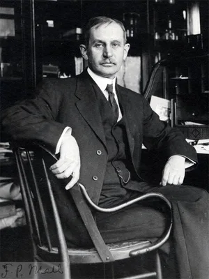
Mall defined the liver's structural unit as a tissue block centered on a portal vein, rather than the classic hepatic lobule centered on a central vein. He proposed that the structural unit of the liver is defined by …
1750 CE
#10122
Anthropotomie, ou L'Art de disséquer les muscles, les ligamens, les nerfs, & les vaisseaux sanguins du corps humain; auquel on a joint une histoire succincte de ces vaisseaux; avec la manière de faire les injections; de préparer, de blanchir les os & de dresser les squelettes. De préparer toutes les différentes parties & de les conserver préparées, soit dans une liqueur propre à cet effet, soit en les faisant sécher; celle d'ouvrir & d'embaumer les cadavres. On y donne aussi la description des matières propres à chacune de ces préparations, & la figure des instrumens. 2 vols.
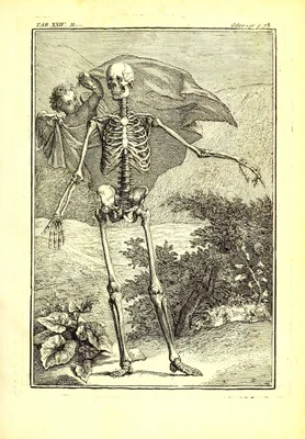
An exceptionally thorough manual on dissection, dressing skeletons, and creating "anatomical preparations," and embalming. Digital facsimile from BnFGallica at this link.
1992 CE
#7354
Brain maps: Structure of the rat brain.
The first computer graphics atlas of the brain of any species, with the illustration files also available separately (1993). The work included a complete and systematic, hierarchically organized set of annotated nomen…
1774 CE
#6835
Kaitai Shinsho (解体新書 Kyūjitai: 解體新書,(Anatomical Tables). A translation of Johan Adam Kulmus's Ontleedkundige Tafelen by Sugita Genpaku.
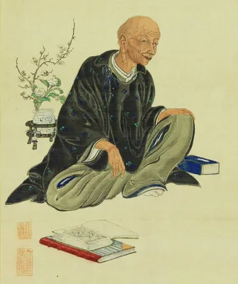
The first translation of any Western medical text into Japanese. "Kaitai Shinsho represented the beginning of two epoch-making developments. First and most directly Gempaku's work set in motion the modern transformati…
1887 CE
#14019
The topographical anatomy of the child.
The first part of this work concerns cross-sectional anatomy; the second part systematically discusses the differences between anatomy in children and adults of different parts of the body.
1991 CE
#11236
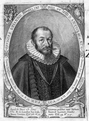
A bibliography of Johann Remmelin the anatomist
1808 CE
#7682
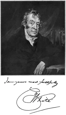
A catalogue of the anatomical preparations, casts, drawings, machines, instruments, &c. in White's Museum, Lying-in hospital.
1831 CE
#768.1
A critical and experimental essay on the circulation of the blood.
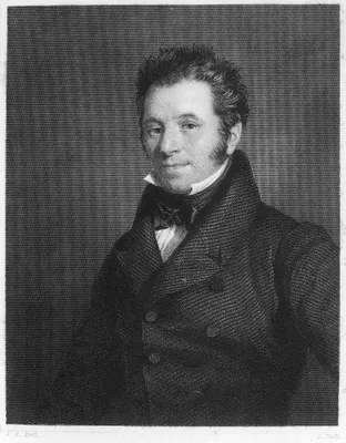
Marshall Hall clearly distinguished arterioles and venules from capillaries, and he described arteriovenous shunts.
1911 CE
#7644
A cross-section anatomy, by Albert C. Eycleshymer and Daniel M. Schoemaker. Average position of organs from eleven reconstructions, by Peter Potter. Sections of the female pelvis, by Carroll Smith. Drawings by Tom Jones.
The historical introduction includes a bibliographical history of cross-sectional anatomies from frozen sections. Digital facsimile from the Hathi Trust at this link.
1788 CE
#399.2
A description of all the bursae mucosae of the human body.
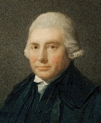
The first serious study of this subject and the most original anatomical work by the greatest of the Monro dynasty. See No. 1385.
1730 CE
#1217
A description of the peritonaeum, and of that part of the membrane cellularis which lies on its outside. With an account of the true situation of all the abdominal viscera, in respect of these two membranes.
Douglas described the peritoneum in detail; his name is perpetuated in the “pouch”, “line”, and “fold of Douglas”. He was a friend of John Hunter and brother of John Douglas, the li…
2003 CE
#9804
A history of online information services 1963-1976.
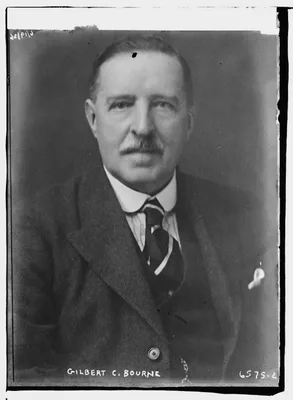
Pages 197-223 concern "Modern bibliographic control of medical literature." Development of MEDLARS, MEDLARS II, MEDLINE.
1852 CE
#415
A manual of artistic anatomy.
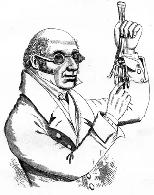
Knox, remembered because of his indiscreet association with the Edinburgh “resurrectionists”, was one of the best teachers of anatomy during the 19th century.
1836 CE–1842 CE
#13299
A series of anatomical plates. 5 vols.: The muscles of the human body. The vessels of the human body. The nerves of the human body. The viscera of the human body. The bones and ligaments of the human body.
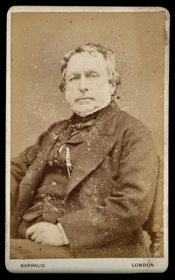
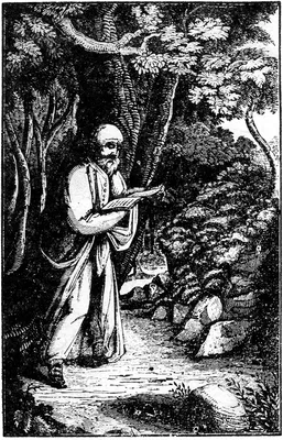
1836 CE–1842 CE
#13300
A series of anatomical plates. 5 vols.: The muscles of the human body. The vessels of the human body. The nerves of the human body. The viscera of the human body. The bones and ligaments of the human body.
The most ambitious 19th century English anatomy illustrated by lithography. Some copies were issued with hand-colored plates. The five volumes, containing a total of 201 plates, describe the muscles, blood vessels, ne…
1732 CE
#393
A syllabus or index, of all the parts that enter the composition of the human body…For the use of those that go through courses of anatomy.
Chovet was born in England and died in Philadelphia. He made many beautiful wax models to illustrate his lectures, and was among the first to popularize the use of wax and natural preparations in the teaching of anato…
1822 CE–1826 CE
#13508
A system of anatomical plates of the human body. By John Lizars. Text in 8vo in 12 pts., plus folio atlas with engraved title and 101 plates engraved by William Home Lizars.
Lizars's System of anatomical plates was by far his most successful work, going through many editions. The text of the work was originally issued in 12 parts in octavo format, which were then bound together in book fo…
1811 CE–1813 CE
#11320
A system of anatomy or the use of students of medicine. 2 vols.
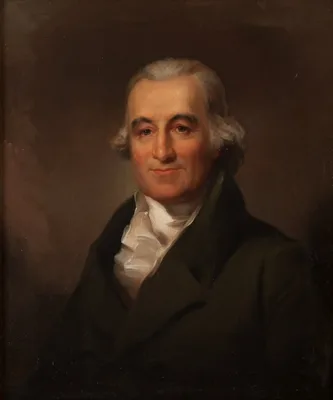
The first American textbook of anatomy. The first edition contained nearly 1000 pages of text, but no illustrations. Later editions were expanded, illustrated and updated by William E. Horner, and Joseph Pancoast. Dig…
1798 CE–1803 CE
#402
A system of dissections. 2 vols.
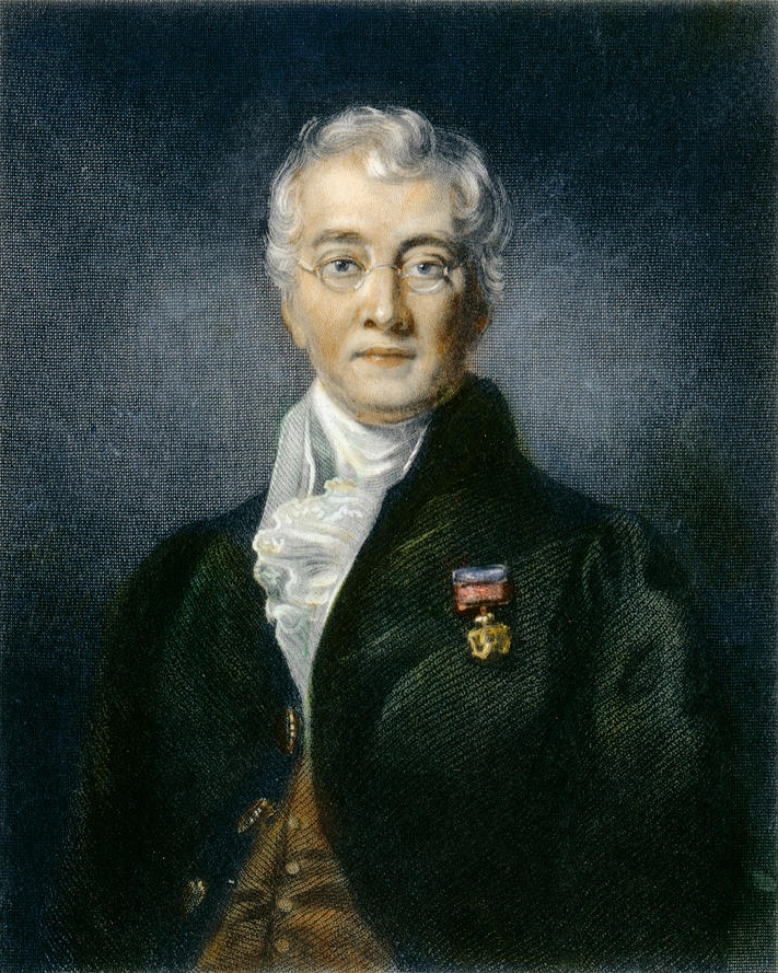
Published in 7 fascicules and appendix while Bell was still a student, this was Bell’s first independent venture as an author. The anatomical work of Charles Bell and his brother John was among the most signific…
1858 CE
#419
A treatise on the human skeleton, including the joints.
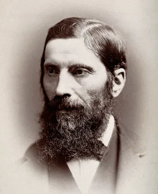
Humphry was professor of anatomy at Cambridge and became the first professor of surgery there. He founded the Journal of Anatomy and Physiology in 1867. “Humphry’s ligament” of the knee-joint is desc…
1829 CE
#11760
Address to the community, on the necessity of legalizing the study of anatomy
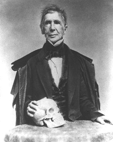
The petition to the Massachusetts legislature to legalize "the procuring of subjects for anatomical dissections" (from George Hayward's printed notice on the verso of the title page). Nine members of the Massachusetts…
1691 CE
#1213
Adenographia curiosa et uteri foeminei anatome nova.
Description of the “canal of Nuck”.
1656 CE
#1116
Adenographia: sive, glandularum totius corporis descriptio.
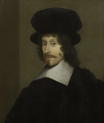
Wharton described the duct of the submaxillary salivary gland (“Wharton’s duct”). He described the thyroid more accurately than his predecessors, naming it. He also described “Wharton’s j…
1818 CE
#408
Afbeeldingen van de juiste plaatsing der inwendige deelen van het menschelijk ligchaam.
First anatomical illustrations of frozen sections. De Riemer appears to have been the first to freeze tissues in order to permit fine sectioning for the purposes of diagnosing diseased tissue. Digital facsimile from U…
1841 CE
#543
Allgemeine Anatomie. Lehre von den Mischungs- und Formbestandtheilen des menschlichen Körpers.
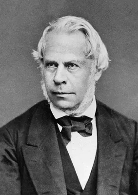
Many of the histological discoveries of Henle are described in the above. He classified tissues histologically. In the section on Gefässnerven (pp. 510, 690) Henle demonstrated the presence of smooth muscle in th…
1919 CE–1922 CE
#10206
American Frohse anatomical charts. Edited, revised and augmented by Max Brödel. With: A key to the Frohse anatomical charts.
10 wall charts, each 42 x 64 inches, comprising a total of 76 colored illustrations life size or larger. Chart 1: Human Skeleton Chart 2: Muscles, front and back Chart 3: Nervous and Circulatory Systems Chart 4: a Sch…
1699 CE
#1214
An account of two new glands and their excretory ducts, lately discovered in human bodies.
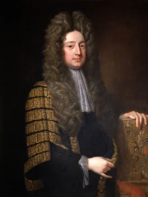
Cowper’s description of the glands which bear his name. He was forestalled in their discovery by Jean Méry.
1849 CE
#13868
An atlas of anatomical plates of the human body, with descriptive letter-press in English and Hindustani.
Digital facsimile from the Internet Archive at this link.
1943 CE
#7347
An atlas of the basal ganglia, brain stem and spinal cord, Based on myelin-stained material.
"Classic atlas of the human brain (excluding the cerebral and cerebellar cortex). Each structure has a blurb with varying amounts of useful historical and factual information. There is also a very useful bibliography …
1861 CE
#421
An elementary treatise on human anatomy.
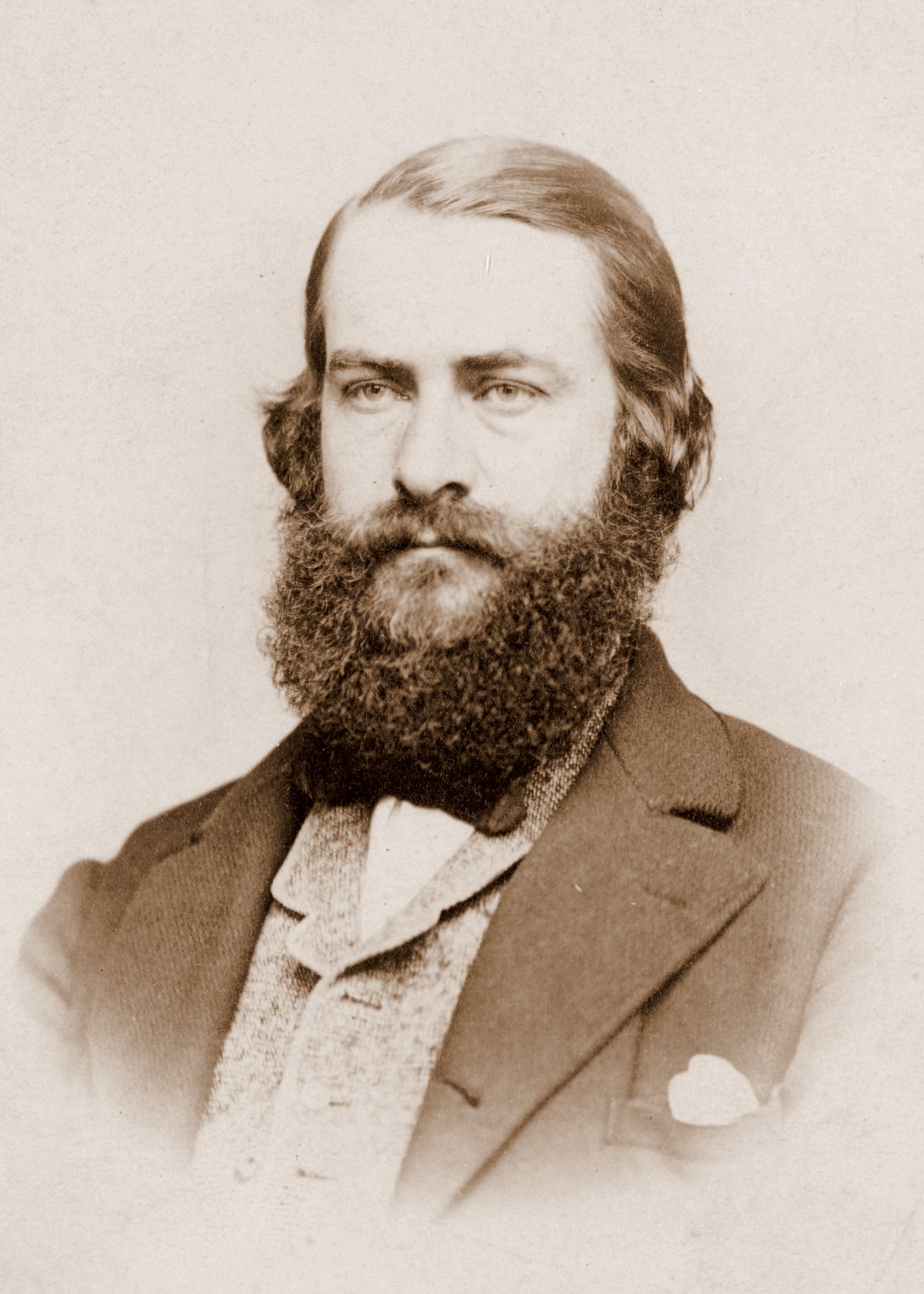
Leidy illustrated this book himself. He was professor of anatomy at Philadelphia and the leading American anatomist of his time.
1756 CE
#7489
An essay on the demonstration of the human structure, half as large as nature, in four tables, from the pictures painted after dissections, for that purpose.
Four large mezzotint plates issued with an accompanying text.
2002 CE
#14288
An introduction to human evolutionary anatomy.

1689 CE
#12782
Anatome ossium novis inventis illustrata.
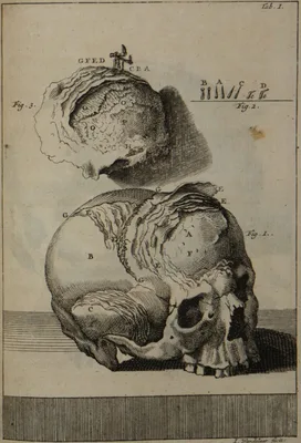
Gagliardi, a papal physician, fiirst described the lamellar structure of the bones. He also carried out some tests and comparative research between human and calf bones, and probably first described a case of bone tub…
1852 CE–1859 CE
#416
Anatome topographica sectionibus per corpus humanum congelatum triplici directione ductis illustrata. 8 pts.
Pirogov was the greatest of Russian surgeons. He introduced the teaching of applied topographical anatomy in Russia. His atlas of 220 plates represents the first use on a grand scale of frozen sections in anatomical i…
1837 CE–1841 CE
#11697
Anatomia chirurgica truncorum arteriarum nec non fasciarum fibrosarum.
An atlas of arterial stems and fasciae. Translated into German as Chirurgische anatomie der Arterienstamme und Fascien neu Bearbeitet von Julius Szymanowski. Leipzig und Heidelberg: C. F. Winter, 1850. Digital facsimi…
1672 CE
#384
Anatomia chirurgica.
First book devoted entirely to surgical anatomy.
1693 CE
#388
Anatomia corporis humani, in qua omnia tam veterum, quam recentiorum anatomicarum inventa methodo nova & intellectu facillima describuntur, ac tabulis aenis repaesentantur.
This work was widely used for some years after publication, superseding Bartholin in popularity. Second edition, with supplement, 2 vols., Louvain, 1706-12.
1739 CE
#13152
Anatomia do corpo humano, recopilada com doutrinas medicas, chimicas, filosoficas, mathematicas, com indices, e estampas, representantes todas as partes do corpo humano.
The first textbook of anatomy published by a Portuguese author in Portuguese. Digital facsimile from Google Books at this link.
1654 CE
#1098.1
Anatomia hepatis … subjiciuntur nonnulla de lymphae-ductibus nuper repertis.
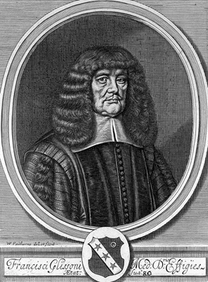
Independently of Bartholin and Rudbeck, George Joyliffe (1621-58) observed the lymphatics. He communicated his discovery to Glisson early in 1652 and the latter included an account in the above work (Cap. xxxi). See N…
1654 CE
#972
Anatomia hepatis.
First accurate description of the capsule of the liver (Glisson’s capsule) and its blood-supply. He also described the sphincter of the bile duct (“Glisson’s sphincter”, the sphincter of Oddi).…
1685 CE
#385
Anatomia humani corporis, centum et quinque tabulis, per artificiosiss. G. de Lairesse ad vivum delineatis.
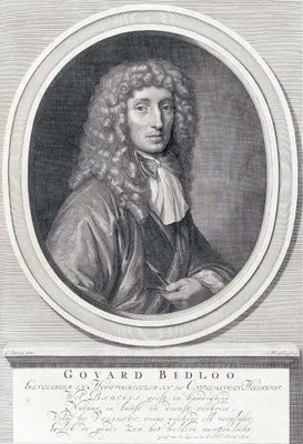
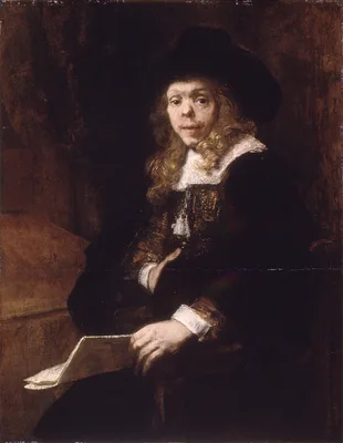
This large folio by Dutch physician, anatomist, poet, and playwright Govert Bidloo contains an engraved title, engraved portrait of Bidloo by Abraham Bloteling after Gérard de Lairesse and 105 engraved plates a…
1811 CE
#406
Anatomia per uso de’pittori e scultori.
This anatomy for artists and sculptors contains 38 good copperplates in black and red.
1816 CE
#7488
Anatomia per uso degli studiosi di scultura e pittura.
Mascagni's anatomy for artists and sculptors, edited for posthumous publication by Mascagni's literary execultor Francesco Antonmarchi. 15 hand-colored engraved plates after drawings by Antonio Serantoni (1780j-1837),…
1691 CE
#386
Anatomia per uso et intelligenza del disegno ricercata non solo su gl’ossi, e muscoli del corpo humano.
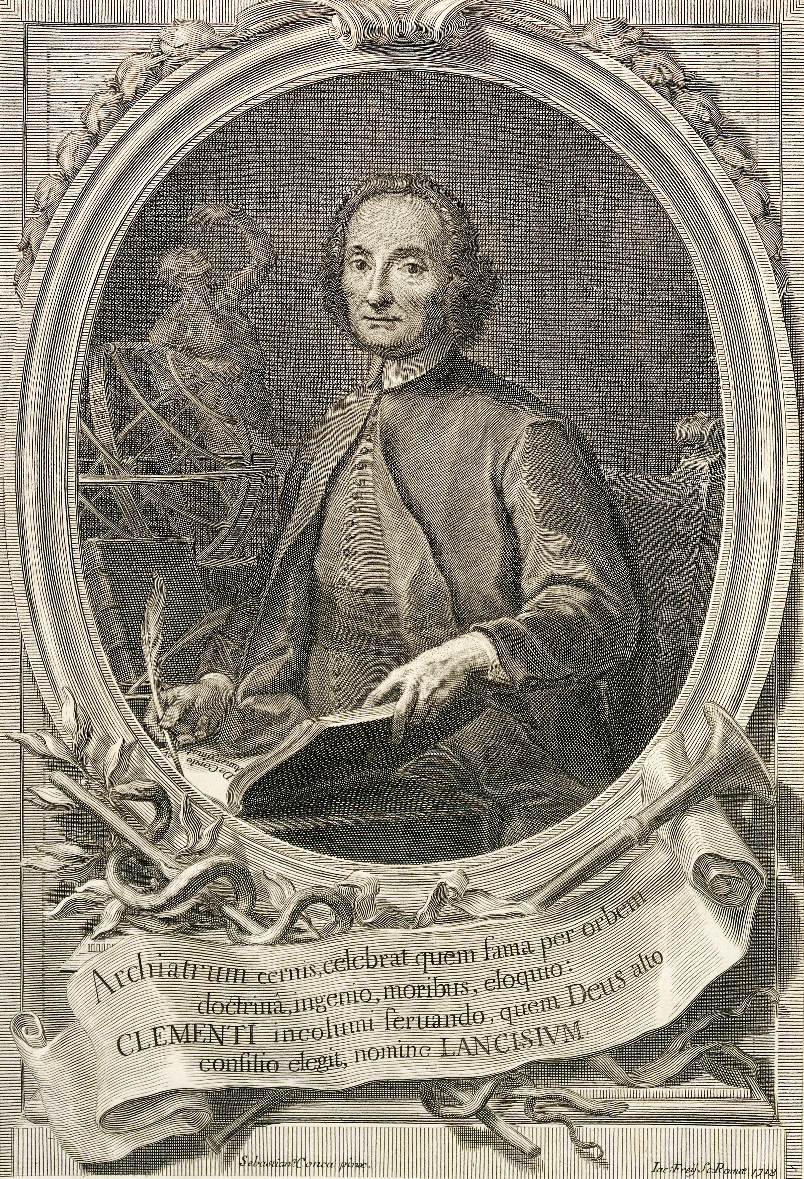
Contains 56 copper-plates, excellent anatomically and artistically, with commentary by Giovanni Maria Lancisi. This is one of the finest of all books on anatomy for artists. English translation with plates re-engraved…
1822 CE–1832 CE
#409.1
Anatomia universa… 2 vols.
The largest of all medical books from the standpoint of format. The 44 life size engraved plates are reproduced in double elephant folio size measuring 950 x 635 mm., and include an almost incredible level of detail. …
1611 CE
#6964
Anatomicae institutiones corporis humani.
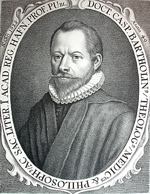
The elder Caspar Bartholin was the first to describe the workings of the olfactory nerve, and introduced the terms nervus olfactorius and nervus vagus. This was a standard textbook for many years, undergoing numerous …
1845 CE
#2294.1
Anatomical and pathological observations.
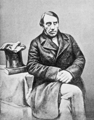
John Goodsir’s paper on “Centres of nutrition” anticipates to a certain extent the cell doctrine afterwards developed by Virchow (see No. 2299). Virchow dedicated the first edition of his Cellularpat…
1833 CE
#411
Anatomical studies of the bones and muscles, for the use of artists. From drawings by the late John Flaxman, Esq. R.A. Engraved by Henry Landseer. With two additional plates, and explanatory notes, by William Robertson.
Digital facsimile from digitalcollections.nypl.org at this link.
1821 CE–1831 CE
#409
Anatomie de l’homme, ou descriptions et figures lithographiées de toutes les parties du corps humain. 5 vols.
The first anatomical atlas illustrated by lithography, containing 300 plates in folio format. This was one of the most elaborate of the lithographic “incunabula” produced by Charles Philibert de Lasteyrie,…
1748 CE
#7491
Anatomie de la tête, en tableaux imprimés qui représentent au naturel le cerveau sous différentes coupes, la distribution des vaisseaux dans toutes les parties de la tête, les organes des sens et une partie de la névrologie, d'après les pièces disséquées et préparées par M. Duverney, en huit grandes planches dessinées, peintes, gravées et imprimées en couleur et grandeur naturelle....
Includes eight spectacular plates printed in color by Gautier d'Agoty with text by Duvereny. Digital facsimile from the Internet Archive at this link.