
Facets
Browse across eight MeSH (opens in new tab) facets — era, geography, science, specialty, technology, history, culture, and reference. Select one tag per group; counts update across the others.
Clear filtersFacet filters
Geography
Specialties & Disease
- Anatomy & Pathology 290
- Cardiology & Blood 3
- Neurology & Psychiatry 19
- Obstetrics & Reproductive 3
- Infectious Disease (General) 0
- Surgery & Anesthesia 14
- Public Health 6
- Immunology & Dermatology 11
- General Clinical Medicine 4
- Military Medicine 0
- Psychology 1
- Alternative & Fringe Medicine 0
- Pediatrics 2
- Ophthalmology & Vision 4
- ENT & Hearing 5
- Urology & Nephrology 12
- Gastroenterology & Hepatology 20
- Pulmonary & Respiratory 1
- Rheumatology, Rehab & Pain 0
- Internal, Emergency & Geriatric 0
- Veterinary Medicine 1
- Epidemiology & Demography 0
- Physiology & Embryology 29
- Dentistry 4
- Plagues & Epidemics 0
- Microbiology & Virology 3
Social & Historical Studies
Institutions & Culture
Reference & Scholarly Works
Drugs & Technology
290 entries match Modern [K01.400.504] · Anatomy & Pathology [G02.149 / C23]
1733 CE
#395
Osteographia, or the anatomy of the bones.
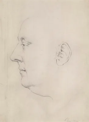
This splendidly designed and illustrated work contained full and accurate descriptions of all the human bones, as well as many of animals. Cheselden is the first person to have used the camera obscura to gain precisio…
1691 CE
#387
Osteologia nova, or some new observations of the bones.
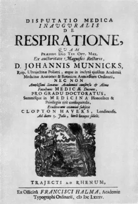
Havers discovered the Haversian canals and made important observations of the physiology of bone growth and repair. The Haversian lamellae, glands, and folds, are also named after him. The Haversian canals were observ…
1828 CE
#7512
Otium academicum, continens descriptionem speciminum nonnullarum partium corporis humani et animalium subtilioris anatomiae ope in physiologicum usum praeparatarum, aliarumque, quibus morborum organicorum natura illustrator.
Issued in 12 fascicules from 1826 to 1828. Consists of 3 parts concerning anatomy and physiology, comparative anatomy specimens and pathological specimens. Includes 37 plates printed in color; 35 plates in black & whi…
1818 CE–1822 CE
#534.57
Philosophie anatomique. Tome Premier. Des organes respiratoires sou le rapport de la détermination et de l'identité de leur pièces osseuses. Avec figures de 116 nouvelles preparations d'anatomie. [Tome Deuxième]. Des monstruosités humaines, ouvrage contenant une classification des monstres; la description et la comparaison des principaux genres; une histoire raisonnée des phénomènes de la monstruosité et des faits primitifs qui la produisent; des vues nouvelles touchant la nutrition du foetus et d'autres circonstrances de son développement....Avec figures des détails anatomiques. 2 vols.
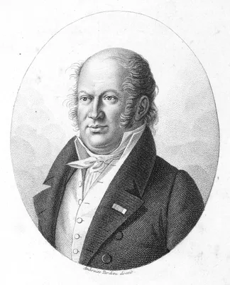
The elder Geoffroy Saint-Hilaire is credited with coining the word teratology, and was the first seriously to attempt the experimental production of anomalies, by manipulating chick eggs. See T. Cahn, La vie et l'oeuv…
1823 CE–1826 CE
#7242
Planches anatomiques du corps humain executes d’après les dimensions naturelles. Double elephant folio atlas and small folio text.
Considering that it is among the rarest of all anatomies, and certainly the largest and probably the most spectacular, it is remarkable that two nearly identical editions of Mascagni’s posthumous life-size anato…
1886 CE
#427
Plastische Anatomie des menschlichen Körpers.
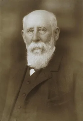
“Illustrated with lithographs from hand-drawings, photographs from the nude, ethnic studies of facial features…The text…is of unusual historic interest, and includes special chapters on the anatomy…
1924 CE
#13680
Plastische Anatomie: Die konstruktive Form des menschlichen Körpers. Mit Bildern von Hermann Sachs.
An anatomy for artists, illustrated by Hermann Sachs.
1891 CE
#1018.1
Quelques points de l’anatomie et de la chirurgie des voies biliaires.
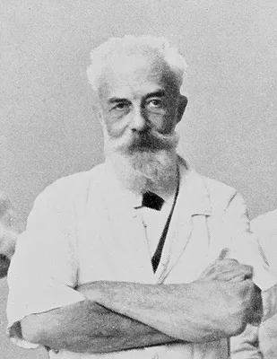
“Hartmann’s pouch”, a dilatation of the neck of the gall-bladder.
1891 CE–1892 CE
#10587
Regional anatomy in its relation to medicine and surgery. 2 vols.
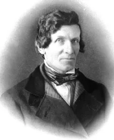
Includes 97 beautiful chromolithographed plates dissected, photographed, and colored from nature by McClellan. Digital facsimile of the 2nd edition from the Internet Archive at this link.
1828 CE
#7096
Report from the select committee on anatomy. House of Commons, 22 July 1828.
In the first half of 1828, in response to increasing calls for reform, the British Parliament appointed a committee to "enquire into the manner of obtaining subjects for dissection by schools of Anatomy and the State …
1831 CE
#11759
Report of the Select Committee of the House of Representatives ... legalizing the study of anatomy.
This was the first law passed in the United States consigning the bodies of those who died in workhouses, hospitals, and similar institutions, the bodies of whom were "unclaimed," to medical schools for dissection. "S…
1784 CE–1790 CE
#77
Sämmtliche kleinere Schriften. 3 vols.
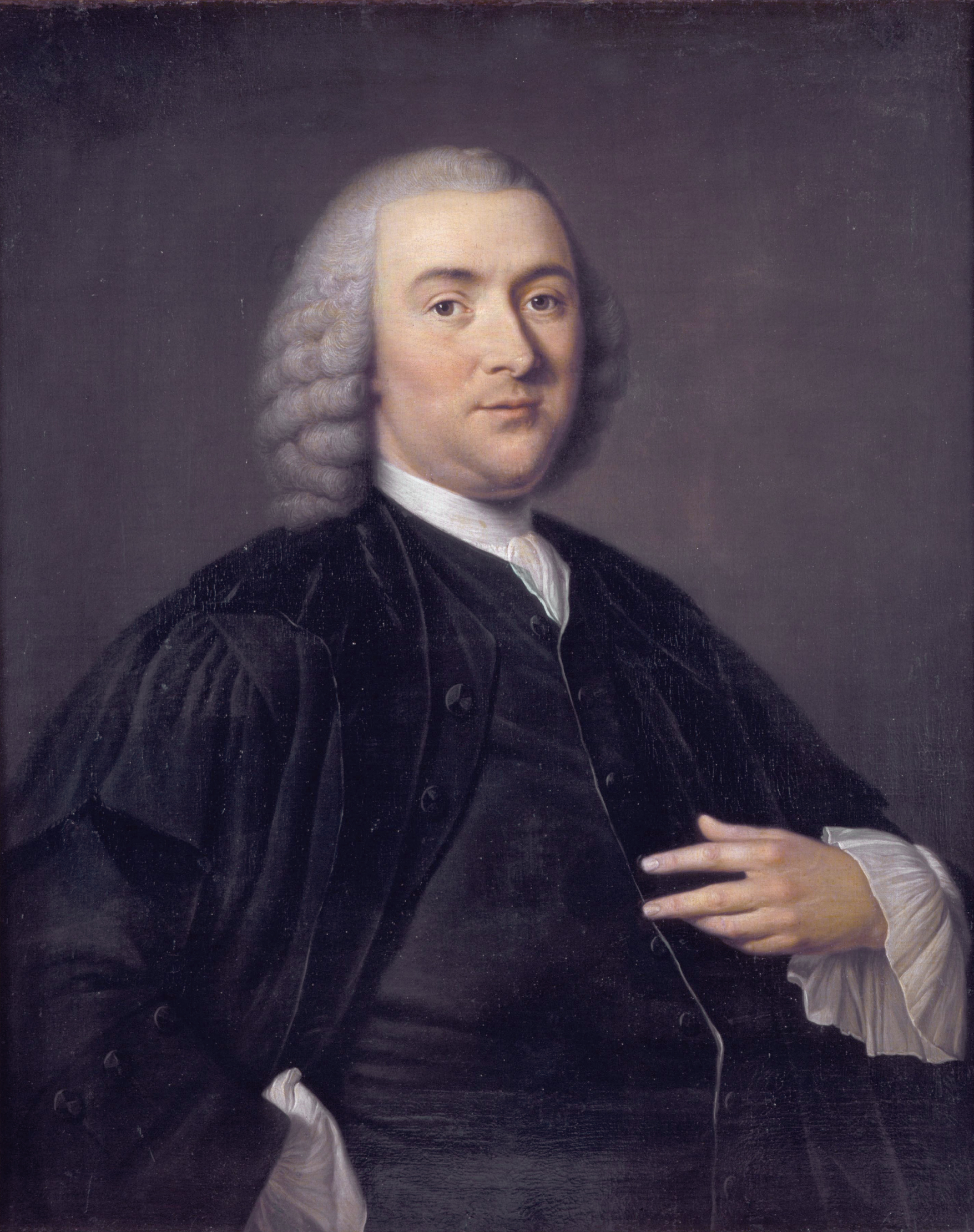
Camper, an artist of skill, made his mark as an anthropologist and craniologist. He discovered the processus vaginalis of the peritoneum and the fibrous structure of the eye, and made several other important contribut…
1775 CE
#399.1
Septemdecim tabulae…
Santorini died before the completion of these anatomical plates which he intended to be his chef d’oeuvre. This elegantly printed volume is the only significant medical book printed by the celebrated Giambattist…
2001 CE
#10707
Skulls and skeletons: Human bone collections and accumulations.
1670 CE
#383
Spicilegium anatomicum.
Kerckring made important investigations on the development of the foetal bones. He was the first to describe the large ossicle sometimes present at the lambdoidal suture; his name is remembered in the valvulae connive…
1851 CE
#13302
Surgical anatomy.
"The drawings of Maclise for Quain's Anatomy of the arteries and for his own Surgical anatomy are indeed done, as Quain wrote, with spirit and effect. These figures of anatomical dissection seem lifelike; in many plat…
1730 CE
#977
Suspenseurs de l’abdomen.
“Poupart’s ligament”, the inguinal ligament.
1742 CE
#396.1
Syndesmologia sive historia ligamentorum corporis humani.
Weitbrecht is known for “Weitbrecht’s ligament” (of the elbow), “Weitbrecht’s foramen ovale” (gap in the capsule of the shoulder joint between the glenohumeral ligaments), and &ldqu…
1641 CE
#9417
Syntagma anatomicum, publicis dissectionibus, in auditorum usum, diligenter aptatum.
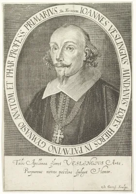
Vesling provided an early discussion of the human lymphatic system. He was one of the first physicians to describe the brain's circle of Willis. Digital facsimile from the Internet Archive at this link.
1822 CE
#13314
System of surgical anatomy. Part first [all published]: On the structure of the groin, pelvis, and perineum. As connected with inguinal and femoral hernia; Tyeing the iliac arteries; and the operation of lithotomy. Illustrated by nine copper-plate engravings
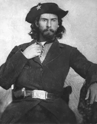
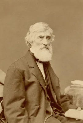
Anderson trained at the Royal College of Surgeons in Edinburgh, emigrated to America in 1820 and, after delivering a series of anatomical lectures in New York under the patronage of Valentine Mott, became affiliated w…
1797 CE
#401
Tabula sceleti feminini juncta descriptione.
Soemmerring was noted for his accuracy in anatomical illustration, and the above work is a fine example of his artistic sense. For it he selected the skeleton of a well-built girl of 20 years. Great care was taken in …
1627 CE
#381
Tabulae anatomicae lxxiix.
First publication of the very beautiful copperplates engraved by Francesco Valesio after Odoardo Fialetti, a pupil of Titian. Casseri commissioned these plates covering the whole field of human anatomy for his unfinis…
1752 CE
#6018
Tabulae anatomicae quatuor uteri duplicis.
Atlas of bipartite and double uterus.
1714 CE
#1312
Tabulae anatomicae.
A romantic history attaches to this fine collection of plates, drawn by Eustachius himself and completed in 1552. They remained unprinted and forgotten in the Vatican Library until discovered in the early 18th century…
1741 CE
#395.2
Tabulae anatomicae.
![PIETRO DA CORTONA, [Pietro BERRETINI] (1596 – 1669)](/media/portraits/743)
27 anatomical copperplates after drawings by the most influential painter of the Italian Baroque movement, who also excelled as an architect. The editor, Cajetano Petrioli, supplied the text and small numbered anatomi…
1737 CE–1747 CE
#399
Tabulae sceleti et musculorum corporis humani.
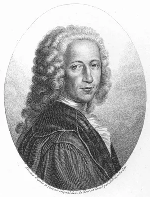
The splendid series of 40 large copperplates of the bones and muscles in this work were drawn and engraved by Jan Wandelaar (1690-1759). They established a newstandard in anatomical illustration, and remain unsurpasse…
1971 CE
#14020
The anatomical basis of medical practice.

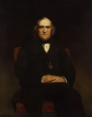
A notorious anatomy textbook that was pulled from the market by the publisher due to protest over its use of Playboy magazine type models to illustrate female external anatomy, as well as its sexist language. Preface:…
1790 CE
#7329
The anatomical instructor; or, an illustration of the modern and most approved methods of preparing and preserving the different parts of the human body, and of quadrupeds, by injection, corrosion, maceration, distention, articulation, modelling, &c.
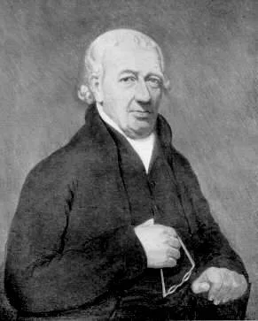
The first monograph on the preparation of anatomical specimens for museums, from various parts of the human body. Includes a method for injecting colored solutions to show the blood vessels of the head, and a method f…
1868 CE
#13221
The anatomical memoirs of John Goodsir F.R.S. Edited by William Turner. With a biographical memoir by Henry Lonsdale. 2 vols.
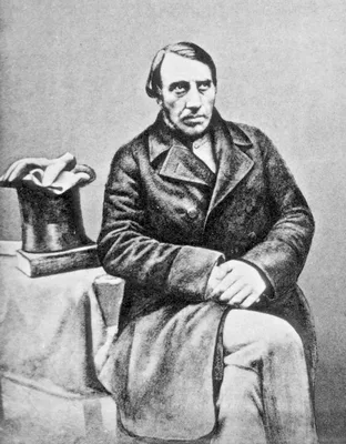
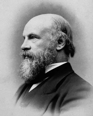
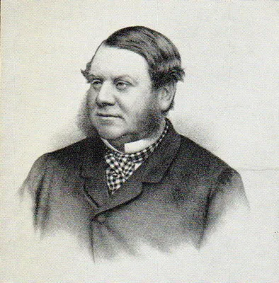
1836 CE
#7674
The anatomist's instructor, and museum companion; being practical directions for the formation and subsequent management of anatomical museums.
"Dr Frederick Knox was the librarian of New Zealand's first public library. He emigrated from Scotland in July 1840 and within days of arriving in Port Nicholson became involved in establishing the country's first pub…
1834 CE
#10024
The anatomy and surgery of inguinal and femoral hernia.
Published in the same large folio format as Tuson's Myology (1828), this was the largest work on hernia ever published with multiple hand-colored. lift-up flats on three plates.
1698 CE
#385.1
The anatomy of humane bodies, with figures drawn after the life by some of the best masters in Europe.
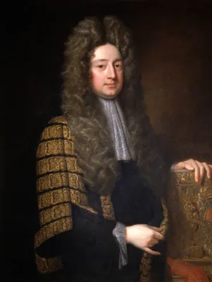
The largest in format, and most elaborate and beautiful of all 17th century English treatises on anatomy, and also one of the most extraordinary plagiarisms in the entire history of medicine. Cowper purchased sets of …
1786 CE
#1103
The anatomy of the absorbing vessels of the human body.
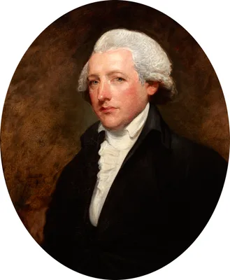
With Hunter and Hewson, Cruikshank laid the foundation of modern knowledge concerning the lymphatics. He was Dr. Johnson’s physician and William Hunter’s assistant.
1797 CE–1804 CE
#401.3
The anatomy of the human body. 4 vols.
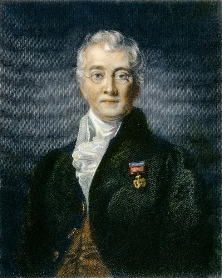
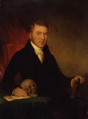
“The first great textbook contributed by the British school to modern anatomy” (Russell, No. 461).
1834 CE
#1497
The anatomy of the human eye.
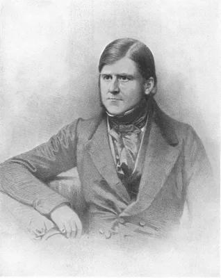
First English work on ocular anatomy.
1713 CE
#390
The anatomy of the humane body.
Although Cheselden is best known for his accomplishments in the field of surgery, he wrote two important books on anatomy. The above was for many years a textbook of the English medical schools and ran through 13 edit…
1986 CE
#14021
The anatomy of the infant head.
1832 CE
#1119
The anatomy of the thymus gland.
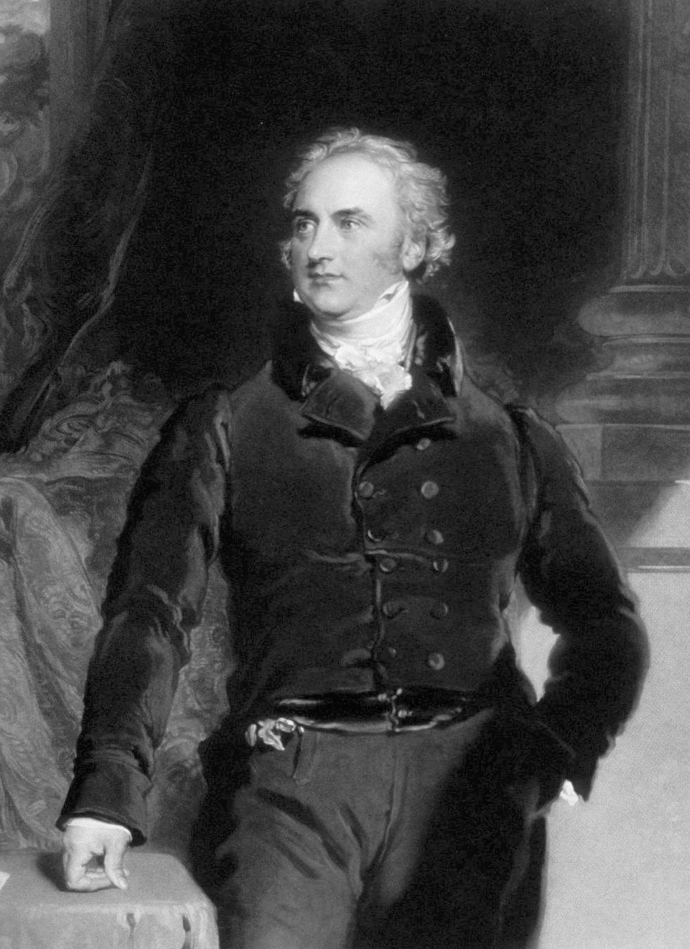
Cooper, the most popular surgeon in London during the early part of the 19th century, was connected with both Guy’s and St. Thomas’s Hospitals. Among his best works is his description of the thymus; he des…
1998 CE
#7350
The central nervous system of vertebrates. 3 vols.
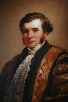
A massive contribution to comparative vertebrate neuroanatomy, the life-work of the authors. Includes a comprehensive account of the structural organisation of all vertebrate groups, ranging from amphioxus and lamprey…
1948 CE
#10204
The Ciba collection of medical illustrations. A compilation of pathological and anatomical paintings prepared by Frank H. Netter, M.D.
This was the first collection of anatomical images by Netter published in book form.
1892 CE
#12786
The collected papers of Sir W. Bowman. Edited by J. Burdon-Sanderson and J. W. Hulke. 2 vols.
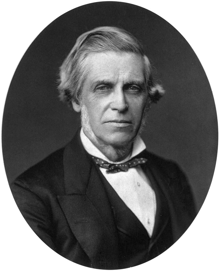
Digital facsimile from Google Books at this link.
1998 CE
#7642
The complete visible human: The complete high-resolution male and female anatomical datasets from the Visible Human Project.
The first anatomically exact and complete, three-dimensional, computer-generated reconstruction of actual human bodies. Includes 2 CD-ROMs. See https://www.nlm.nih.gov/research/visible/visible_human.html.
1835 CE–1859 CE
#603
The cyclopaedia of anatomy and physiology. Edited by Robert Bentley Todd. 5 vols.
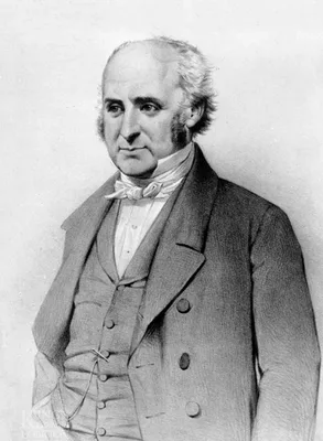
Contributors included Richard Owen and Thomas Huxley, and physicians James Paget, James Young Simpson, and William Bowman.The discoveries of Purkynĕ and Valentin, together with additional observations by William Sharp…
1905 CE–1906 CE
#10599
The Edinburgh stereoscopic atlas of Anatomy. Edited by David Waterston. 5 vols.
The first large scale application of stereoscopic photography to anatomy. The 5 parts each contain 50 stereo cards on which are pasted original stereo photographs and corresponding printed descriptive text. Digital fa…
2012 CE
#12143
The evolution of the human placenta.
Chapter one is "The history of placental investigations."
2009 CE
#11313
The finger of God: Anatomical practice in seventeenth-century Leiden.
1864 CE
#7331
The gray substance of the medulla oblongata and trapezium.
The first American medical book illustrated with photomechanically reproduced plates. Oliver Wendell Holmes praised the book for its remarkable photomicrographs, which may be the first published of brain cross-section…
1833 CE
#411.1
The hand: Its mechanism and vital endowments as evincing design.
Classic work on the anatomy, physiology, bio-mechanics, comparative anatomy, and adaptive importance of the hand. Issued as a volume in a series entitled the "Bridgewater Treatises." The first edition has 288pp. An en…
1959 CE
#13256
The human body, what it is and how it works. Text by Mitchell Wilson. Illustrations by Cornelius De Witt. Arthur W. Seligmann, M.D., medical consultant.
A modern classic of medical illustration, and the popularization of medicine. The artist is best known for illustrating children's books.
1954 CE
#7351
The human brain in sagittal section.
A superb atlas is based on sagittal sections. This was an innovative approach for the time as almost all previous illustration of the adult human brain was typically based on frontal or horizontal sections: “in …