
Facets
Browse across eight MeSH (opens in new tab) facets — era, geography, science, specialty, technology, history, culture, and reference. Select one tag per group; counts update across the others.
Clear filtersFacet filters
Geography
Specialties & Disease
- Anatomy & Pathology 79
- Cardiology & Blood 1
- Neurology & Psychiatry 4
- Obstetrics & Reproductive 1
- Infectious Disease (General) 0
- Surgery & Anesthesia 5
- Public Health 1
- Immunology & Dermatology 0
- General Clinical Medicine 1
- Military Medicine 0
- Psychology 0
- Alternative & Fringe Medicine 0
- Pediatrics 0
- Ophthalmology & Vision 0
- ENT & Hearing 0
- Urology & Nephrology 1
- Gastroenterology & Hepatology 0
- Pulmonary & Respiratory 0
- Rheumatology, Rehab & Pain 0
- Internal, Emergency & Geriatric 0
- Veterinary Medicine 1
- Epidemiology & Demography 0
- Physiology & Embryology 5
- Dentistry 2
- Plagues & Epidemics 0
- Microbiology & Virology 0
Social & Historical Studies
Institutions & Culture
Reference & Scholarly Works
Drugs & Technology
79 entries match Modern [K01.400.504] · Anatomy & Pathology [G02.149 / C23] · Arts, Literature & Humanities [K01.090]
1992 CE
#7354
Brain maps: Structure of the rat brain.
The first computer graphics atlas of the brain of any species, with the illustration files also available separately (1993). The work included a complete and systematic, hierarchically organized set of annotated nomen…
1852 CE
#415
A manual of artistic anatomy.
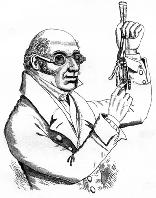
Knox, remembered because of his indiscreet association with the Edinburgh “resurrectionists”, was one of the best teachers of anatomy during the 19th century.
1836 CE–1842 CE
#13300
A series of anatomical plates. 5 vols.: The muscles of the human body. The vessels of the human body. The nerves of the human body. The viscera of the human body. The bones and ligaments of the human body.
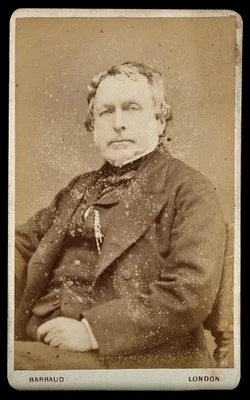
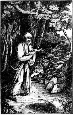
The most ambitious 19th century English anatomy illustrated by lithography. Some copies were issued with hand-colored plates. The five volumes, containing a total of 201 plates, describe the muscles, blood vessels, ne…
1822 CE–1826 CE
#13508
A system of anatomical plates of the human body. By John Lizars. Text in 8vo in 12 pts., plus folio atlas with engraved title and 101 plates engraved by William Home Lizars.
Lizars's System of anatomical plates was by far his most successful work, going through many editions. The text of the work was originally issued in 12 parts in octavo format, which were then bound together in book fo…
1798 CE–1803 CE
#402
A system of dissections. 2 vols.
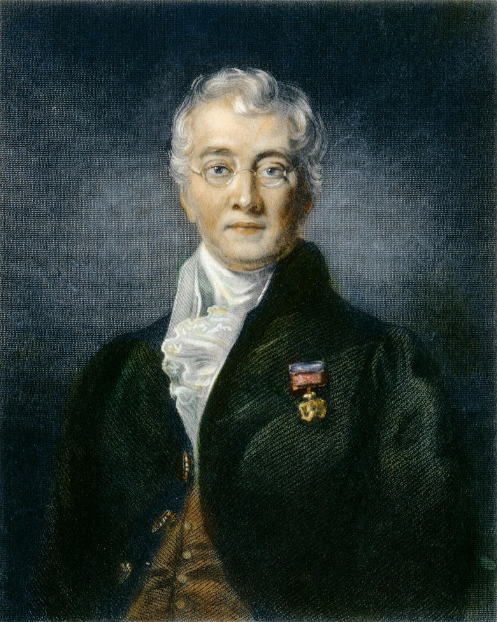
Published in 7 fascicules and appendix while Bell was still a student, this was Bell’s first independent venture as an author. The anatomical work of Charles Bell and his brother John was among the most signific…
1818 CE
#408
Afbeeldingen van de juiste plaatsing der inwendige deelen van het menschelijk ligchaam.
First anatomical illustrations of frozen sections. De Riemer appears to have been the first to freeze tissues in order to permit fine sectioning for the purposes of diagnosing diseased tissue. Digital facsimile from U…
1756 CE
#7489
An essay on the demonstration of the human structure, half as large as nature, in four tables, from the pictures painted after dissections, for that purpose.
Four large mezzotint plates issued with an accompanying text.
1852 CE–1859 CE
#416
Anatome topographica sectionibus per corpus humanum congelatum triplici directione ductis illustrata. 8 pts.
Pirogov was the greatest of Russian surgeons. He introduced the teaching of applied topographical anatomy in Russia. His atlas of 220 plates represents the first use on a grand scale of frozen sections in anatomical i…
1693 CE
#388
Anatomia corporis humani, in qua omnia tam veterum, quam recentiorum anatomicarum inventa methodo nova & intellectu facillima describuntur, ac tabulis aenis repaesentantur.
This work was widely used for some years after publication, superseding Bartholin in popularity. Second edition, with supplement, 2 vols., Louvain, 1706-12.
1685 CE
#385
Anatomia humani corporis, centum et quinque tabulis, per artificiosiss. G. de Lairesse ad vivum delineatis.
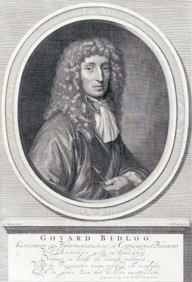
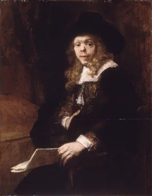
This large folio by Dutch physician, anatomist, poet, and playwright Govert Bidloo contains an engraved title, engraved portrait of Bidloo by Abraham Bloteling after Gérard de Lairesse and 105 engraved plates a…
1811 CE
#406
Anatomia per uso de’pittori e scultori.
This anatomy for artists and sculptors contains 38 good copperplates in black and red.
1816 CE
#7488
Anatomia per uso degli studiosi di scultura e pittura.
Mascagni's anatomy for artists and sculptors, edited for posthumous publication by Mascagni's literary execultor Francesco Antonmarchi. 15 hand-colored engraved plates after drawings by Antonio Serantoni (1780j-1837),…
1691 CE
#386
Anatomia per uso et intelligenza del disegno ricercata non solo su gl’ossi, e muscoli del corpo humano.
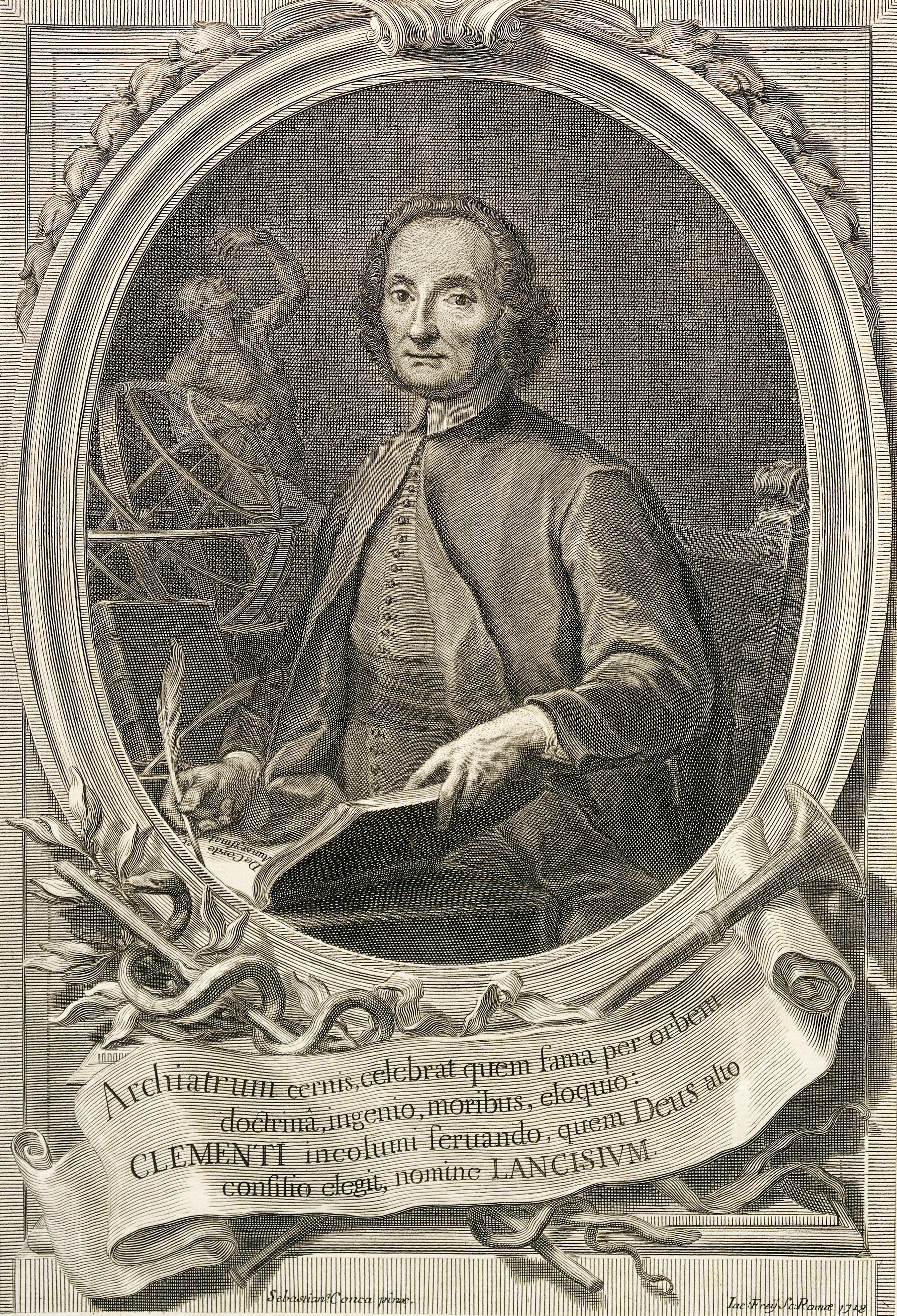
Contains 56 copper-plates, excellent anatomically and artistically, with commentary by Giovanni Maria Lancisi. This is one of the finest of all books on anatomy for artists. English translation with plates re-engraved…
1822 CE–1832 CE
#409.1
Anatomia universa… 2 vols.
The largest of all medical books from the standpoint of format. The 44 life size engraved plates are reproduced in double elephant folio size measuring 950 x 635 mm., and include an almost incredible level of detail. …
1833 CE
#411
Anatomical studies of the bones and muscles, for the use of artists. From drawings by the late John Flaxman, Esq. R.A. Engraved by Henry Landseer. With two additional plates, and explanatory notes, by William Robertson.
Digital facsimile from digitalcollections.nypl.org at this link.
1821 CE–1831 CE
#409
Anatomie de l’homme, ou descriptions et figures lithographiées de toutes les parties du corps humain. 5 vols.
The first anatomical atlas illustrated by lithography, containing 300 plates in folio format. This was one of the most elaborate of the lithographic “incunabula” produced by Charles Philibert de Lasteyrie,…
1748 CE
#7491
Anatomie de la tête, en tableaux imprimés qui représentent au naturel le cerveau sous différentes coupes, la distribution des vaisseaux dans toutes les parties de la tête, les organes des sens et une partie de la névrologie, d'après les pièces disséquées et préparées par M. Duverney, en huit grandes planches dessinées, peintes, gravées et imprimées en couleur et grandeur naturelle....
Includes eight spectacular plates printed in color by Gautier d'Agoty with text by Duvereny. Digital facsimile from the Internet Archive at this link.
1634 CE
#381.1
Anatomie der uuterlicke deelen van het menschelick lichaem: Dienende om te verstaen ende volkometlick wt te beelden alle beroerlicheit des selven lichaems.
The earliest of all independent works on anatomy for graphic or plastic artists. The author, a painter and etcher, drew and engraved all the images himself. Digital facsimile from Google Books at this link.
1829 CE
#11162
Anatomie des formes extérieures du corps humain appliquée à la peinture, à la sculpture et à la chirurgie.
Digital facsimile from the Internet Archive at this link.
1812 CE
#14179
Anatomie du gladiateur combattant, applicable aux beaux arts . . . .
Salvage's 21 plates after his own drawings "are based on three casts of bodies dissected to different anatomical layers and set in the pose of the Borghese Gladiator. For these casts he preferred to use the bodies of …
1754 CE
#7492
Anatomie generale des visceres, et de la nevrologie, angeologie et osteologie du corps humain, en figures, de couleurs et grandeurs naturelles dediée et présentée.
Includes 18 full-page color-printed mezzotints, 12 of the plates designed to fit together in threes to make four life-size human figures. Gautier credited Mertrud, the King's Surgeon with some of the anatomical work i…
1995 CE
#11142
Anatomy atlases: An anatomy digital library curated by Ronald A. Bergman.
https://www.anatomyatlases.org/ "About Us Curate a comprehensive digital library of anatomy information for patients and providers. Maximize the impact of this digital library by enhancing awareness among potential us…
1945 CE
#9177
Anatomy for artists.
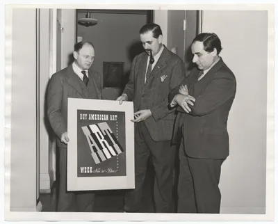
By the famous American social realist artist.
1900 CE
#10588
Anatomy in its relation to art. An exposition of the bones and muscles of the human body with especial reference to their influence upon its actions and external form.
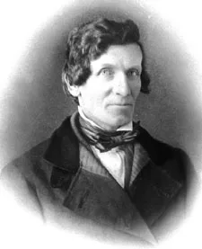
The work was offered for sale by J. B. Lippincott with title pages dated 1901. Digital facsimile of the Lippincott issue from Google Books at this link.
1858 CE
#418
Anatomy, descriptive and surgical
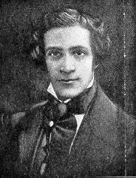
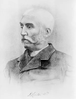
Gray’s textbook of anatomy remains today a standard work on the subject in the English-speaking world. The 37th edition appeared in 1989; the first American edition was published at Philadelphia, 1859. Digital f…
1989 CE
#436.1
Atlas of human anatomy.
The culmination of the life work of one of the greatest, and most prolific, anatomical illustrators of the 20th century. Includes 514 full-color plates, many of which were created for this atlas. Reproduction of previ…
2002 CE
#10238
BioDigital.
https://www.biodigital.com/ "The Word's First Human Visualization Platform: Anatomy, Disease & Treatments— all in interactive 3D. Web, Mobile and Augmented Reality "the virtual body as the health equivalent of G…
1613 CE
#11237
Catoptri micorcosmici absolutam admirandae partium hominis creaturarum divinarum praestantissimi fabricae eximio artificio sculptam structuram revidendam exhibentis. Engraved by Stephan Michelspacher.
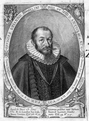
Three large anatomical plates with numerous flaps, in the tradition of fugitive sheets, but larger and more complex. The first edition was issued anonymously without identifying the place of publication or the publish…
1953 CE–1990 CE
#10203
Ciba collection of medical illustrations. 8 vols. in 13.
"In all, Netter produced nearly 4,000 illustrations, which have been included in countless publications. In perspective, that number represents an image researched, sketched, and completely painted for every three bus…
1651 CE
#382
Corporis humani disquisitio anatomica.
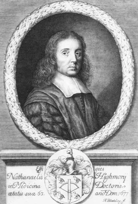
Highmore is remembered for his description of the maxillary sinus, known eponymically as the “antrum of Highmore” (already noticed by Casserius and figured by Leonardo da Vinci), the seminal ducts and the …
1922 CE–1931 CE
#7649
Das Leben des Menschen. Eine volkstümliche Anatomie, Biologie, Physiologie und Entwicklungsgeschichte des Menschen. 5 vols.
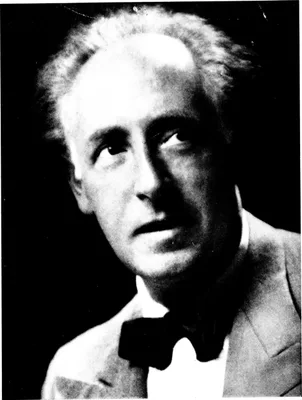
By developing a new infographics style of illustration in which physiological processes and other technical medical and biological concepts were often depicted as, or compared to machines, Kahn made medical and biolog…
1884 CE–1889 CE
#784
Das Venensystem des menschlichen Körpers. 2 pts. and atlas.
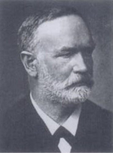
Like Braune’s other anatomical works, this is notable for its excellent illustrations.
1611 CE
#380
De anatome corporis humani libri vii.
Guidi, professor of philosophy and medicine at Pisa, discovered the Vidian nerve, the Vidian canal, and the Vidian artery. The above was edited by his nephew.
1627 CE
#1094
De lactibus sive lacteis venis.
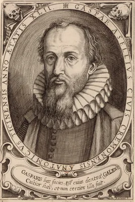
Records the discovery of the lacteal vessels. While performing vivisection on a dog that had recently fed, Aselli noticed a network of vessels in the mesentery and along the peritoneal surface of the intestine. The ve…
1736 CE
#7507
Dissertatio de arteries et venis intestinorum hominis. Adjecta icon coloribus distincta.
The small color mezzotint printed by the painter Jan Ladmiral included with this pamphlet on the arteries and veins of the human intestine was among the earliest applications of full color printing, and the first use …
1745 CE
#398
Essai d’anatomie en tableaux imprimés, qui represent au naturel tous les muscles de la face, du col, de la tête, de la langue & du larinx. d'après les parties disséquées & préparées par Monsieur Duverney....comprenant hit grandes planches.
Remarkable for its striking mezzotints printed in color. Digital facsimile from the Internet Archive at this link.
2011 CE
#7005
Fragonard Museum. The écorchés. The anatomical masterworks of Honoré Fragonard by Christophe Degueurce. With an essay by Laure Cadot. Translated from the French by Philip Adds.
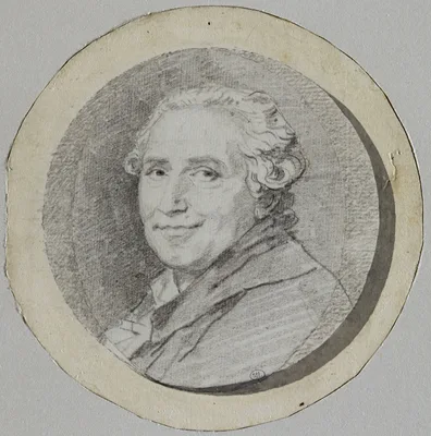
The painter and printmaker Fragonard preserved the results of his dissections via means never divulged, but which may have been based on those of Jean-Joseph Sue. His pieces were often prepared for theatrical effect r…
1734 CE
#7552
Historia musculorum hominis.
Very detailed descriptions of all the muscles of the human body, with illustrations drawn and engraved by Jan Wandelaar depicting the muscles of the hand, life-size with all the muscles, tendons, ligaments, and bones.…
1743 CE–1756 CE
#397
Icones anatomicae. 8 pts.
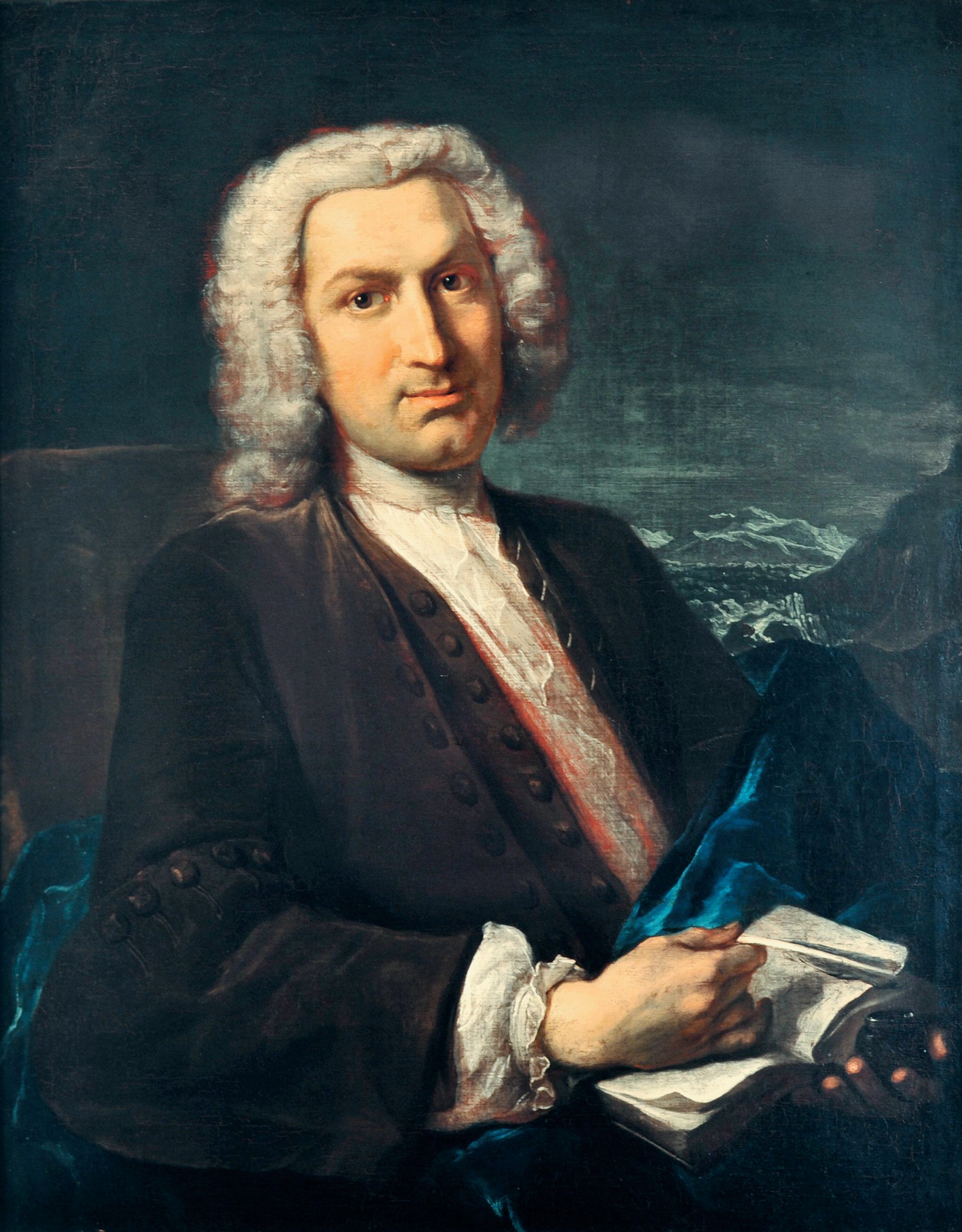
Accurate and beautiful engravings of the diaphragm, uterus, ovaries, vagina, arteries, with explanatory observations. About fifty years after they were originally published the most visually spectacular versions of Ha…
1934 CE
#6622.1
John Keats’s anatomical and physiological note book …edited by Maurice Buxton Forman.

Keats was a pupil and dresser at Guy’s Hospital from 1815-16, and was licensed to practice upon completion of his studies. While struggling to launch his poetic career he was often tempted to practice medicine, …
1813 CE
#13214
Kaitai hatsumo [Explanation of Human Anatomy]. 5 vols.
The first Japanese exposition of the whole system of human anatomy, based on original observation. The work includes the records of three dissections performed in Kyoto in 1783, 1798, and 1802. “A Japanese treat…
1991 CE
#10202
Max Brödel, The man who put art into medicine
In the late 1890s, Brödel was brought to the Johns Hopkins School of Medicine to illustrate for Harvey Cushing, William Halsted, Howard Kelly, and other notable clinicians. Besides creating a prolific amount of w…
2019 CE
#10777
Medicine and religion in the life of an Ottoman sheikh: Al-Damanhuri's "clear statement" on anatomy.
1828 CE
#10023
Myology, illustrated by plates.
This large folio work contains 8 hand-colored lithographed plates by F. R. Say, each with multiple lift-up flaps. It is probably the largest format anatomical work ever published in English with lift-up flaps, with up…
1724 CE
#392.1
Myotomia reformata.
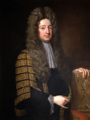
This work made a modest first appearance in 1694 as an octavo, but Cowper worked until his death on a new edition which was finally published posthumously under the supervision and at the expense of Richard Mead (1673…
1684 CE
#1379
Neurographia universalis.
Vieussens, professor at Montpellier, was the first to describe the centrum ovale correctly. The publication of the above work threw new light on the subject of the configuration and structure of the brain, spinal cord…
2000 CE
#7643
New atlas of human anatomy.
The first printed atlas of color computer images adapted from 3D images developed in the National Library of Medicine's Visible Human Project. Includes CD-ROM with 3D electronic images.
1779 CE
#401.1
Nouveau recueil d’ostéologie et de myologie…
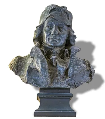
“Without contest the most beautiful of all anatomies for the artist and one of the most remarkable books of its time” (Hahn & Dumaitre). The plates in this work are more fantastic than any other anatomy, s…
1725 CE
#8095
Opera omnia anatomica & chirurgica. Edited by Herman Boerhaave and Bernhard Siegfried Albinus. 2 vols.
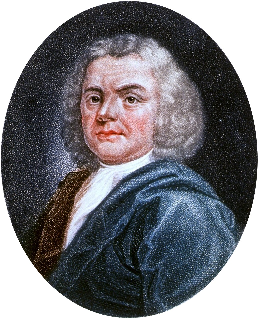
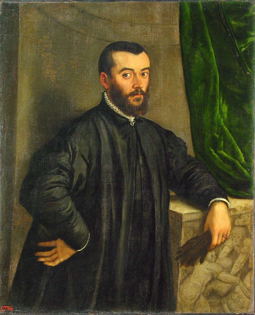
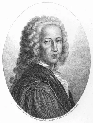
Vesalius's collected works with the famous woodcuts reproduced as copperplate engravings by Jan Wandelaar (1690-1759). Notably Boerhaave and Albinus had this edition published because Vesalius's works still had practi…
1645 CE
#61.2
Opera quae extant omnia. Ex recension Joh. Antonidae vander Linden.
Spieghel succeeded Casseri in the chair of anatomy at Padua. This edition of his collected writings contains the second printing of the 97 copperplates first printed in Casseri’s Tabulae anatomicae (No. 381) plu…